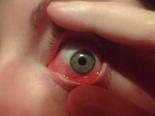
Conjunctivitis (also called pink eye or madras eye) refers to inflammation of the conjunctiva (the outermost layer of the eye and the inner surface of the eyelids). It is most commonly due to an infection (usually viral, but sometimes bacterial) or an allergic reaction.
By cause
- Allergic conjunctivitis
- Bacterial conjunctivitis
- Viral conjunctivitis
- Chemical conjunctivitis
- Neonatal conjunctivitis is often defined separately due to different organisms
By extent of involvement
Blepharoconjunctivitis is the dual combination of conjunctivitis with blepharitis (inflammation of the eyelids).
Keratoconjunctivitis is the combination of conjunctivitis and keratitis (corneal inflammation).
Episcleritis is an inflammatory condition that produces a similar appearance to conjunctivitis, but without discharge or tearing.
Signs and symptoms
Red eye (hyperaemia), irritation (chemosis) and watering (epiphora) of the eyes are symptoms common to all forms of conjunctivitis. However, the pupils should be normally reactive and the visual acuity normal.
Viral
Viral conjunctivitis is often associated with an infection of the upper respiratory tract, a common cold, and/or a sore throat. Its symptoms include watery discharge and variable itch. The infection usually begins with one eye, but may spread easily to the other.
Viral conjunctivitis, commonly known as "pink eye", shows a fine, diffuse pinkness of the conjunctiva, which is easily mistaken for the ciliary injection of iritis, but there are usually corroborative signs on microscopy, particularly numerous lymphoid follicles on the tarsal conjunctiva, and sometimes a punctate keratitis.
Bacterial
Bacterial conjunctivitis due to common pyogenic (pus-producing) bacteria causes marked grittiness/irritation and a stringy, opaque, greyish or yellowish mucopurulent discharge that may cause the lids to stick together, especially after sleep. Another symptom that could be caused by bacterial conjunctivitis is severe crusting of the infected eye and the surrounding skin. However, contrary to popular belief, discharge is not essential to the diagnosis. Bacteria such as Chlamydia trachomatis or Moraxella can cause a non-exudative but persistent conjunctivitis without much redness. The gritty and/or scratchy feeling is sometimes localized enough for patients to insist they must have a foreign body in the eye. The more acute pyogenic infections can be painful. Like viral conjunctivitis, it usually affects only one eye but may spread easily to the other eye. However, it is dormant in the eye for three days before the patient shows signs of symptoms.Corynebacterium diphtheriaecauses membrane formation in conjunctiva of non immunized children and very rare disease called Acute membrane conjunctivitis
Chemical
Chemical eye injury is due to either an acidic or alkali substance getting in the eye. Alkalis are typically worse than acidic burns. Mild burns will produce conjunctivitis while more severe burns may cause the cornea to turn white. Litmus paper is an easy way to rule out the diagnosis by verifying that the pH is within the normal range of 7.0—7.2. Large volumes of irrigation is the treatment of choice and should continue until the pH is 6—8. Local anaesthetic eye drops can be used to decrease the pain.
Irritant or toxic conjunctivitis show primarily marked redness. If due to splash injury, it is often present only in the lower conjunctival sac. With some chemicals, above all with caustic alkalis such assodium hydroxide, there may be necrosis of the conjunctiva with a deceptively white eye due to vascular closure, followed by sloughing of the dead epithelium. This is likely to be associated with slit-lamp evidence of anterior uveitis.
Other
Inclusion conjunctivitis of the newborn (ICN) is a conjunctivitis that may be caused by the bacteria Chlamydia trachomatis, and may lead to acute, purulent conjunctivitis. However, it is usually self-healing.
Conjunctivitis is identified by irritation and redness of the conjunctiva. Except in obvious pyogenic or toxic/chemical conjunctivitis, a slit lamp (biomicroscope) is needed to have any confidence in the diagnosis. Examination of the tarsal conjunctiva is usually more diagnostic than the bulbar conjunctiva.
Causes
Conjunctivitis is most commonly caused by viral infection, but bacterial infections, allergies, other irritants and dryness are also common etiologies for its occurrence. Both bacterial and viral infections are contagious. Commonly, conjunctival infections are passed from person-to-person, but can also spread through contaminated objects or water.
The most common cause of viral conjunctivitis is adenoviruses. Herpetic keratoconjunctivitis (caused by herpes simplex viruses) can be serious and requires treatment with acyclovir. Acute hemorrhagic conjunctivitis is a highly contagious disease caused by one of two enteroviruses, Enterovirus 70 and Coxsackievirus A24. These were first identified in an outbreak in Ghana in 1969, and have spread worldwide since then, causing several epidemics.
Diagnosis
Cultures are done infrequently because most cases of conjunctivitis are treated empirically and (eventually) successfully, but often only after running the gamut of the common possibilities.
Swabs for bacterial culture are necessary if the history and signs suggest bacterial conjunctivitis, but there is no response to topical antibiotics. Research studies indicate many bacteria implicated in low-grade conjunctivitis are not detected by the usual culture methods of medical microbiology labs, so false negative results are common. Viral culture may be appropriate in epidemic case clusters. Conjunctival scrapes for cytology can be useful in detectingchlamydial and fungal infections, allergy and dysplasia, but are rarely done because of the cost and the general lack of laboratory staff experienced in handling ocular specimens. Conjunctival incisional biopsy is occasionally done whengranulomatous diseases (e.g., sarcoidosis) or dysplasia are suspected.
Pink Eye Relief products....


No comments:
Post a Comment